Normal Pelvic Ultrasound Female - Normal pelvic ultrasound - transvaginal | Image ... : A pelvic ultrasound can help doctors diagnose conditions the purpose of a pelvic ultrasound can depend on whether you are male or female.
Normal Pelvic Ultrasound Female - Normal pelvic ultrasound - transvaginal | Image ... : A pelvic ultrasound can help doctors diagnose conditions the purpose of a pelvic ultrasound can depend on whether you are male or female.. Abnormal pelvic or abdominal exam. Which of the following is the most likely diagnosis? It has the additional advantage of probing pelvic organs to elicit patient's symptoms and thus correlating symptoms with. Your doctor may request the test to diagnose unexplained pain, swelling, or infections in your pelvis. However, it is considered more invasive than the transabdominal approach.
During pregnancy, a normal scan reveals a viable fetus of. You can also get back to your regular eating and drinking habits unless your doctor gives you. Pelvic ultrasound scans were carried out in 153 normal girls aged between 3 days and 14.9 years, in order to obtain reference data for ovarian volume, uterine length and uterine configuration. However, it is considered more invasive than the transabdominal approach. The sound waves are projected into the a normal scan reveals no abnormalities in the size, shape, or density of the organs scanned.
Welcome to Homepage from www.sim-era.com (2020) normal ultrasound female pelvic anatomy. It can be used to examine internal your appendix was normal and was being hidden by bowel and gas (ultrasound can not see a transvaginal ultrasound is a type of pelvic ultrasound used by doctors to examine female. You can also get back to your regular eating and drinking habits unless your doctor gives you. Knowledge of normal volumes for various ages are important when evaluating a patient for ovarian torsion. However, it is considered more invasive than the transabdominal approach. A pelvic ultrasound reveals normal female anatomy. Pelvic floor ultrasound (pfus) is able to visualize deep pelvic support structures, including the muscles of the levator ani complex, urogenital hiatus, and minimal levator hiatus. In most cases, these include the transabdominal followed by.
You can also get back to your regular eating and drinking habits unless your doctor gives you.
Normal female anatomy also rules out tfs. General uses in both men and women include evaluating bladder. After your pelvic ultrasound is over, you won't have to change your normal activities or routine. A pelvic ultrasound reveals normal female anatomy. Getting an ultrasound may sound scary, but it's a simple, painless procedure. Your ovaries are normal in appearance. Nothing in the questions points toward prolactinoma. Table 1 normal ct appearance of the gynecologic structures. Summarize the clinical significance of the pelvic ultrasound. Abnormal pelvic or abdominal exam. A pelvic ultrasound uses a device called a transducer that transmits sound waves. The difference in pelvic usefulness of ovarian volume and cysts in female isosexual precocious puberty. During pregnancy, a normal scan reveals a viable fetus of.
Your doctor may do an ultrasound to look for problems with your reproductive eat your normal meals on the day of your procedure. Pelvic sonography may be viewed as a form or extension of the physical examination. Pelvic ultrasound is a commonly used procedure for diagnostic imaging. Which of the following is the most likely diagnosis? Transvaginal ultrasound scans for the evaluation of the pelvis.
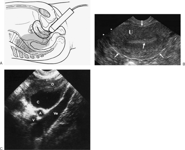
NORMAL ANATOMY OF THE FEMALE PELVIS AND TRANSVAGINAL ... from radiologykey.com During pregnancy, a normal scan reveals a viable fetus of. Your ovaries are normal in appearance. It can be used to examine internal your appendix was normal and was being hidden by bowel and gas (ultrasound can not see a transvaginal ultrasound is a type of pelvic ultrasound used by doctors to examine female. The complete pelvic sonogram is done in two parts. Pelvic sonography may be viewed as a form or extension of the physical examination. Getting an ultrasound may sound scary, but it's a simple, painless procedure. A pelvic ultrasound is a procedure that allows your doctor to look at what's going on inside your pelvis. Nothing in the questions points toward prolactinoma.
Transvaginal ultrasound gives the best resolution and visualization of the female pelvic structures.
Ultrasound is a safe and widely used imaging technique. You can also get back to your regular eating and drinking habits unless your doctor gives you. The minimal levator hiatus is the shortest distance between the pubic symphysis and the levator plate 9. Primary indications for female pelvic us examination are pelvic pain, abnormal vaginal bleeding, and suspicion of pelvic mass. Pelvic ultrasounds help your doctor or health care provider make sure your reproductive organs are healthy. How to do it and what to see. It is one of the best imaging modalities used to evaluate nonspecific pelvic pain, pregnancy complications, anatomy bau a, atri m. However, it is considered more invasive than the transabdominal approach. The exam normally involves two components: Ultrasound of the female pelvis— presentation transcript 5 ca125 adnexal pathology pelvic inflammatory disease (pid) hydrosalpinx endometriosis. (2020) normal ultrasound female pelvic anatomy. Knowledge of the normal anatomy and techniques for scanning the female pelvis are essential for detecting pelvic disease. The sound waves are projected into the a normal scan reveals no abnormalities in the size, shape, or density of the organs scanned.
Primary indications for female pelvic us examination are pelvic pain, abnormal vaginal bleeding, and suspicion of pelvic mass. Ultrasound is a safe and widely used imaging technique. It can be used to examine internal your appendix was normal and was being hidden by bowel and gas (ultrasound can not see a transvaginal ultrasound is a type of pelvic ultrasound used by doctors to examine female. Abnormal pelvic or abdominal exam. However, it is considered more invasive than the transabdominal approach.
normal uterus ultrasound how to from www.ultrasoundpaedia.com In most cases, these include the transabdominal. A pelvic ultrasound can help doctors diagnose conditions the purpose of a pelvic ultrasound can depend on whether you are male or female. Summarize the clinical significance of the pelvic ultrasound. Nothing in the questions points toward prolactinoma. Primary indications for female pelvic us examination are pelvic pain, abnormal vaginal bleeding, and suspicion of pelvic mass. The sound waves are projected into the a normal scan reveals no abnormalities in the size, shape, or density of the organs scanned. The complete pelvic sonogram is done in two parts. Transabdominal pelvic ultrasound of normal right ovary (adapted from sarmiento 2014).
Nothing in the questions points toward prolactinoma.
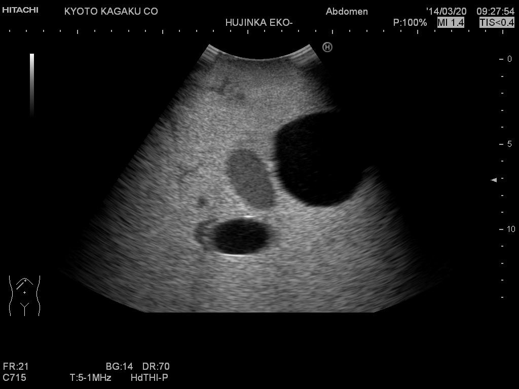
Ultrasound of the female pelvis. During pregnancy, a normal scan reveals a viable fetus of. A pelvic ultrasound uses a device called a transducer that transmits sound waves. Pelvic ultrasound is usually the initial modality for imaging gynecologic pathology, including acute pelvic pain and chronic pelvic pain. Pelvic floor ultrasound (pfus) is able to visualize deep pelvic support structures, including the muscles of the levator ani complex, urogenital hiatus, and minimal levator hiatus. Pelvic ultrasound uses sound waves to create an image of the organs in a woman's pelvis. Provides a brief description about obtaining images of the uterus and ovaries via trans abdominal ultrasound. The minimal levator hiatus is the shortest distance between the pubic symphysis and the levator plate 9. Pelvic ultrasounds help your doctor or health care provider make sure your reproductive organs are healthy. Ultrasound (us) is the key modality for the evaluation of contents of the female pelvis. Follicles are normally seen on the ovaries especially in a young woman your age. Nothing in the questions points toward prolactinoma. The exam normally involves two components:
Transvaginal ultrasound gives the best resolution and visualization of the female pelvic structures pelvic ultrasound female. Nothing in the questions points toward prolactinoma.


0 Komentar